Showing posts with label journal club. Show all posts

Monday, March 1, 2010

Journal Club Follow-Up: Coenzyme Q10

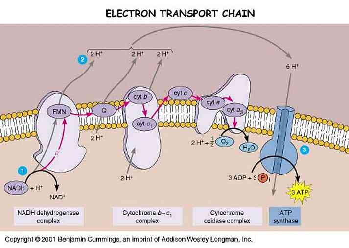
Many Parkinson's patients take Coenzyme Q10 supplements.  As mentioned in the previous post, CoQ10 is part of the Electron Transport Chain -- a very important part, in fact, as it alleviates pressure on our precarious and susceptible-to-aging Complex I.

While many theorize that Complex I is shut down or is deficient in PD (1, 2, 3, 4), others believe that deficient activity of the CoQ10 pool beside Complex I is more to blame (5, 6).  The CoQ10 theory claims that PD causes a deficiency in the CoQ10 pool that carries electrons from Complex I to their next destination without producing ROS.  As a result of low CoQ10, electrons build up in Complex I and get released from the entrance because they cannot leave through the exit.

Some PD patients are able to take CoQ10 supplements and improve their condition (7).

It is my opinion that CoQ10 is a palliative treatment and not a long-term solution.  The Ndi1gene therapy discussed in the previous post is a better option if it makes it to, and proves robust in clinical trials.  My reasoning is that a genetic replacement for Complex I is a more stable therapy than a persistent aid to CoQ10: it is more permanent and a more widespread solution; a large portion of PD patients do not have CoQ10 deficiencies.  Ndi1 would also contribute to the sustaining of the proton gradient in the mitochondria, also vital to creating energy in the ETC.

ResearchBlogging.org
Greenamyre, J. (2001). Response: Parkinson's disease, pesticides and mitochondrial dysfunction Trends in Neurosciences, 24 (5) DOI: 10.1016/S0166-2236(00)01788-4

Schapira AH (1994). Evidence for mitochondrial dysfunction in Parkinson's disease--a critical appraisal. Movement disorders : official journal of the Movement Disorder Society, 9 (2), 125-38 PMID: 8196673

Morais, V., Verstreken, P., Roethig, A., Smet, J., Snellinx, A., Vanbrabant, M., Haddad, D., Frezza, C., Mandemakers, W., Vogt-Weisenhorn, D., Van Coster, R., Wurst, W., Scorrano, L., & De Strooper, B. (2009). Parkinson's disease mutations in PINK1 result in decreased Complex I activity and deficient synaptic function EMBO Molecular Medicine, 1 (2), 99-111 DOI: 10.1002/emmm.200900006

Storch, A., Jost, W., Vieregge, P., Spiegel, J., Greulich, W., Durner, J., Muller, T., Kupsch, A., Henningsen, H., Oertel, W., Fuchs, G., Kuhn, W., Niklowitz, P., Koch, R., Herting, B., Reichmann, H., & , . (2007). Randomized, Double-blind, Placebo-Controlled Trial on Symptomatic Effects of Coenzyme Q10 in Parkinson Disease Archives of Neurology, 64 (7), 938-944 DOI: 10.1001/archneur.64.7.nct60005

Saturday, February 27, 2010

Journal Club: Neuroprotection by NGI1 Gene in a Parkinson's Disease Model

One of the most popular mechanisms of pathology in Parkinson's disease (PD) research is cell death through Complex I inhibition in the mitochondria.

Mitochondria -- affectionately known as the power houses of all cells -- are where energy is produced.  There is a series of protein complexes forming the Electron Transport Chain (ETC) which, as their acronym exposes, steal electrons from contributing molecules and convert them into energy and water.

The fully reduced H2O form of oxygen is non-toxic.  The various single-electron intermediates between O2 and H2O are ALL toxic free radicals, the so-called Reactive Oxygen Species (ROS).  Complex IV (cytochrome oxidase complex) has a gate that transfers electrons directly to O2, reducing it to water without generating ROS.  Complexes I and III, however, occasionally allow electrons to escape from the ETC to form ROS.

In Parkinson's disease, Complex I is dysfunctional, and it is thought that much higher concentrations of ROS are produced in the ETC.  These attack multiple systems in the mitochondria which eventually lead to the breakdown of cellular DNA, and the cell itself.

Many cases of PD are characterized post mortem by a selective loss of dopamine cells via this mechanism.

The study by Marella et al has used the variant of Complex 1 found in yeast to attempt to quell this rampant ROS formation.  Using the rotenone rat model, the group injected the Ndi1 gene via biodegradable microspheres (classy...), and monitored recovery in the substantia nigra pars compacta (SNpc; the primary region of dopamine cell loss) and in behavior.

After 60 days, tissue analysis of Ndi1-injected rotenone rats showed increased staining for viable dopamine cells in the SNpc.  Those lesioned rats who did not receive the Ndi1 gene showed significantly fewer stained dopamine cells, and extensive staining with antibody against 8-oxo-dG (indicating oxidative damage to DNA).

My gripe with the study -- in addition to its not being tremendously written -- is that it is lacking in relevant behavioral assessment.  The study monitored speed of movement, and the number of rotations in a widely used apomorphine test.  The rotations test is normally used in unilaterally lesioned animals (which these were) to indicate preference to rotate in one direction.  However, the direction of rotation induced by apomorphine in this study was determined by more factors than the unilateral lesion, which caused the animals to rotate in both directions.  Therefore, behavioral data was reported as the "number of animals exhibiting 100% lateralized rotation irrespective of the direction."  In my opinion, the behavioral test was severely weakened by this caveat and the group should have employed a quick additional test... like the Whisker test or lateralized grip strength.

This suggests that the Ndi1 gene -- the yeast version of Complex 1 -- was able to compensate for inhibition of Complex 1 by rotenone, decrease ROS activity by serving as an electron transporter, and lessen cell death.  If this could be replicated in higher animals, it may prove a viable candidate for clinical trials. 

Aside from deficits in writing and behavioral analysis, the story told by this article was fascinating with  very intriguing implications.   They did their homework, publishing several studies on in vitro activity of the Ndi1 gene and subsequent protein (1, 2) as well as confirming benign effects of introducing a yeast gene in vivo (1, 2)  .


ResearchBlogging.org
Marella M, Seo BB, Nakamaru-Ogiso E, Greenamyre JT, Matsuno-Yagi A, & Yagi T (2008). Protection by the NDI1 gene against neurodegeneration in a rotenone rat model of Parkinson's disease. PloS one, 3 (1) PMID: 18197244

Sunday, January 24, 2010

Journal Club: RBD and Parkinson's Disease

There is a great deal of research being done regarding the mechanisms of Parkinson's disease (PD) and possible targets for therapeutic cures.  Yet, it is one of many conditions that remains incredibly hard to diagnose.  PD patients are not typically diagnosed until the disease has progressed to 70-90% dopamine cell depletion when symptoms become observable in movement behaviors (Jankovic 2008).

By the time cell loss has progressed this far, it is very difficult to achieve a successful long-term treatment plan.  Pharmaceuticals such as L-Dopa (Jubalt et al 2009) and rasagiline (Olanow et al 2009) are generally effective, but can lose their effect or cause dangerous side effects over time.  Deep brain stimulation has been shown to be very effective behaviorally, but there it is an intense procedure which has occasionally been correlated with subsequent cognitive impairments (York et al 2008).  Exercise therapies have also shown promise in recovery therapy, but have seemed more lasting in the peripheral nervous system than the dopamine system of the CNS (Goodwin et al 2009; Petzinger et al 2007; Muhlack et al 2007).

When it is so important to try to identify markers of PD before it progresses beyond our current ability to treat it in a lasting way, Dr. Ronald Postuma and colleagues out of Montreal, Quebec, Canada have identified REM sleep behavior disorder (RBD) as a possible indication of developing PD.  RBD is the loss of muscle atonia that normally occurs during REM sleep, causing a person to thrash unconsciously.

Their study is a beautiful longitudinal representation of several patients diagnosed with RBD in the 1980s who developed either PD or dementia by 2004.  Of their 17 final RBD patients, 6 (5m/1f) had developed PD and 11 (10m/1f) developed dementia.

The Postuma group suggests that there might be a discrete pathological condition specific to "RBD-then-neurodegeneration"which has different early manifestations than PD alone.  A very interesting concept as RBD, dementia and PD are all distinctive in their Lewy body and ß-amyloid
deposition.  If further study of the evolution of RBD into PD shows a strong correlation, this could be a giant leap forward in terms of PD diagnosis and early treatment.  There may indeed be a distinct pathology to this progression or there may not be.  In any case, this is a very important study in the field of neurodegenerative disorders, and I believe it is expecially important to get longitudinal studies like this one funded.

The staging model of PD developmnt proposed by Braak et al in 2003 proposes that the effects of PD begin in the olfactory area of the brain, spreading to autonomic and sleep-involved regions, and finally to dopamine loss in the nigrostriatal pathway and several downstream cortical pathways (Braak et al 2003).  The Braak model, in conjunction with this new proposal from Postuma et al, leaves me wondering about Restless Leg Syndrome (RLS) as another possible indicator of PD.

The connection between RLS and PD is in dopaminergic transmission, as suggested by Dr. David Rye in 2004.  A study by Tan et al in 2002 found that prevalence of RLS in PD patients was not significantly different from incidence in their healthy population, roughly 15%.  The Tan study was not looking at progression of RLS into PD, however, so it is possible, as suggested in the Postuma study, that RLS-PD may have its own unique pathology. 

To date, I have not found any longitudinal studies of RLS progressing into RBD or PD.

Monday, November 16, 2009

Journal Club: on the selective degeneration of dopamine neurons in Parkinson's disease

http://www.ncbi.nlm.nih.gov/pubmed/16299504

The therapeutic application of potassium gated ATP channels (K-ATP) in Parkinson's disease arises from their ubiquitous expression in the basal ganglia.  Regulation of these channels evokes cell hyperpolarization in order to prevent cell excitability.  In the mitrochondria, they play a role in translating the metabolic state of the neuron.  This week's journal club discussed an article suggesting that K-ATP channels are necessary for the selective vulnerability of dopamine neurons in the substantia nigra pars compacta (SNpc) relative to the ventral tegmental area (VTA).   Liss et al demonstrate this theory using mitrochondrial complex I inhibitors rotenone and MPP+, both neurotoxins commonly used in developing Parkinson's disease models in rodents.

Rotenone and MPP+ are known to selectively degenerate dopamine neurons of the SNpc, leaving the VTA dopamine neurons primarily in tact.  Liss et al suggest that this phenomenon is due to differential mitochondrial uncoupling (or, disruption of metabolism).  Extensive uncoupling with the application of FCCP resulted in activation of K-ATP channels in both the SNpc and VTA.  Mild uncoupling with FCCP did not activate K-ATP channels in either region.
"Notably, however, mild uncoupling inverted the response of K-ATP channels to complex I inhibition: in this case, VTA DA neurons, but not SN DA neurons, were hyperpolarized and functionally silenced due to K-ATP channel activation. In the presence of 50 nM FCCP, none of the SN DA neurons was significantly affected by 100 nM rotenone (Fig. 5a,b, left; perforatedpatch recording in 50 nM FCCP: 2.33 ± 0.29 Hz; FCCP + rotenone: 1.92 ± 0.36, n ¼ 6; P ¼ 0.40) or 10 mM MPP+ (data not shown). In contrast, the presence of 50 nM FCCP sensitized K-ATP channels of VTA DA neurons to complex I inhibition (Fig. 5a,b, right; 50 nM FCCP: 2.4 ± 0.55 Hz; FCCP + rotenone: 0 ± 0 Hz, n ¼ 6; P ¼ 0.0075)."
"Stereological analysis of all SN pars compacta neurons in hematoxylin-eosin counterstained sections demonstrated genuineMPTP-induced neuronal death in wildtype mice and confirmed the complete rescue of SN neurons in the Kir6.2-/- mice (Fig. 6d, middle panel; Kir6.2+/+ SN: control, 11,882 ± 222; post MPTP, 8,061 ± 632, P ¼ 0.029; Kir6.2 -/- SN: control, 12,288 ± 231; post-MPTP, 12,619 ± 223; P ¼ 0.36; n ¼ 3 each)." ** Kir6.2 -/- mice are a genetic strain not expressing a unit of the K-ATP channel necessary for activation.  This means that blocking the channel's activity prevented SN DA neurons from being lost.
I want to see some apoptosis markers in these SNpc DA neurons due to K-ATP activity.  The comaprison of SNpc and VTA DA neurons is an invaluable resource for identifying mechanisms of the selective degeneration that marks Parkinson's disease.  Because the VTA DA neuron population is so identifiably unaffected by most neurotoxins from which Parkinson's models are developed, the selectivity of the models and the degree of neural degeneration is not only measurable but comparable to many cellular mechanisms of the disease itself.  Uncoupling of the mitochondria speaks to selective metabolic toxicity, and a new target for neuroprotective therapies.

** This was a very complex article using six different mouse strains/treatment groups and analyzing the cell viability using electrophysiology, histology and RT-PCR -- I am reciting only the briefest summary which does not to justice to the extensive work done (although my critique is long-winded, I was impressed with these studies).