Showing posts with label protein. Show all posts

Saturday, February 27, 2010

Journal Club: Neuroprotection by NGI1 Gene in a Parkinson's Disease Model

One of the most popular mechanisms of pathology in Parkinson's disease (PD) research is cell death through Complex I inhibition in the mitochondria.

Mitochondria -- affectionately known as the power houses of all cells -- are where energy is produced.  There is a series of protein complexes forming the Electron Transport Chain (ETC) which, as their acronym exposes, steal electrons from contributing molecules and convert them into energy and water.

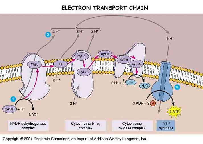
The fully reduced H2O form of oxygen is non-toxic.  The various single-electron intermediates between O2 and H2O are ALL toxic free radicals, the so-called Reactive Oxygen Species (ROS).  Complex IV (cytochrome oxidase complex) has a gate that transfers electrons directly to O2, reducing it to water without generating ROS.  Complexes I and III, however, occasionally allow electrons to escape from the ETC to form ROS.

In Parkinson's disease, Complex I is dysfunctional, and it is thought that much higher concentrations of ROS are produced in the ETC.  These attack multiple systems in the mitochondria which eventually lead to the breakdown of cellular DNA, and the cell itself.

Many cases of PD are characterized post mortem by a selective loss of dopamine cells via this mechanism.

The study by Marella et al has used the variant of Complex 1 found in yeast to attempt to quell this rampant ROS formation.  Using the rotenone rat model, the group injected the Ndi1 gene via biodegradable microspheres (classy...), and monitored recovery in the substantia nigra pars compacta (SNpc; the primary region of dopamine cell loss) and in behavior.

After 60 days, tissue analysis of Ndi1-injected rotenone rats showed increased staining for viable dopamine cells in the SNpc.  Those lesioned rats who did not receive the Ndi1 gene showed significantly fewer stained dopamine cells, and extensive staining with antibody against 8-oxo-dG (indicating oxidative damage to DNA).

My gripe with the study -- in addition to its not being tremendously written -- is that it is lacking in relevant behavioral assessment.  The study monitored speed of movement, and the number of rotations in a widely used apomorphine test.  The rotations test is normally used in unilaterally lesioned animals (which these were) to indicate preference to rotate in one direction.  However, the direction of rotation induced by apomorphine in this study was determined by more factors than the unilateral lesion, which caused the animals to rotate in both directions.  Therefore, behavioral data was reported as the "number of animals exhibiting 100% lateralized rotation irrespective of the direction."  In my opinion, the behavioral test was severely weakened by this caveat and the group should have employed a quick additional test... like the Whisker test or lateralized grip strength.

This suggests that the Ndi1 gene -- the yeast version of Complex 1 -- was able to compensate for inhibition of Complex 1 by rotenone, decrease ROS activity by serving as an electron transporter, and lessen cell death.  If this could be replicated in higher animals, it may prove a viable candidate for clinical trials. 

Aside from deficits in writing and behavioral analysis, the story told by this article was fascinating with  very intriguing implications.   They did their homework, publishing several studies on in vitro activity of the Ndi1 gene and subsequent protein (1, 2) as well as confirming benign effects of introducing a yeast gene in vivo (1, 2)  .


ResearchBlogging.org
Marella M, Seo BB, Nakamaru-Ogiso E, Greenamyre JT, Matsuno-Yagi A, & Yagi T (2008). Protection by the NDI1 gene against neurodegeneration in a rotenone rat model of Parkinson's disease. PloS one, 3 (1) PMID: 18197244

Sunday, August 3, 2008

electron tunneling

In case you were wondering what gets me off...

" By comparison of the measured IETS (*inelastic electron tunneling spectroscopy) spectrum of this molecule with the computed one, it is possible to examine the relative intensities of the different vibrational normal modes, thereby to deduce the pathway for transport. We find that the electrons are injected through the terminal methyl group, tunnel through the sigma bridge to the etheric linkage, mix with the pi electrons, pi tunnel through the aromatic, and switch back to the sigma tunneling, through the thiol and out onto the counter electrode (Galperin and Ratner)."


Bit of background... this was a studying looking at molecular transport junctions wherein a molecule is placed between two electrodes and subjected to applied voltage. This kind of analysis shows particular modes of transport pathways for electrons across the molecule by way of interpreting the molecular junction geometry. This is hot shit because delocalization of electrons across molecules - and in particular, proteins - has the potential to give a great deal of information about protein folding mechanisms...


Protein folding is a fairly simple concept: when DNA copies, it translates its code into a sequence of amino acids. The aa sequence comprises what is called the primary protein structure. AA then fold, through several types of electromagnetic interaction, into secondary structure which begins to take on 3D characteristics. Secondary structure can then fold into several different types of final form, depending on the protein's destined function. But for the purpose of where I'm going with this, I'm going to refer only to primary and secondary structure.

there's a reason that computer modeling has trouble accurately replicating the speed in which proteins fold into their final conformation. that reason is electron tunneling.

in proteins there have been found to exist very significant long-distance tunneling currents of superposed paths over which electrons can delocalize across an entire structure - not just between neighboring atoms which was the conventional assumption.

what this means is that an electron localized to... atom A... can enter into a more delocalized, higher energy superposition between atoms A and B. when it yields back this high energy state, the electron can then relocalize to B rather than back to A, if this is energetically favorable. sound familiar? this is basic electron transfer. now imagine the same mechanism occurring between amino acids of primary and secondary protein structures. because the distance between aa is so much greater than between individual atoms, electron coupling falls away and electrons must delocalize via tunneling currents, connected by "bridges" which enhance tunneling along multiple superposed paths within the molecule.

for instance, in the tubulin protein alpha and beta dimers that make of microtubules (previous post) contain a somewhat patterned arrangement of tryptophan amino acids, arranged all within 2 nanometers of each other. because tryptophan aa contain not only an aromatic ring, but a double aromatic ring (an indole), they provide a tremendously stable delocalization site for electron density, and are thought to be key players in the tunneling current in and between tubulin dimers [this is the tunneling that causes the conformational change in alpha and beta monomers that result in the propagation of an electrical signal down their lattice in the microtubule].

the signal is not just propagated by means of the tryptophan current network; there is also a lattice composes of the aromatic substituents of histidine and phenylalanine amino acids, which correspond to an additional three tunneling possible tunneling patterns. this current-strengthening effect is responsible for the nature of the electromagnetic signal as it is transfered down a microtubule, and from one microtubule to another.

it works as such: the transfer of an electron from one space in a primary structure to another space causes a conformational shift which directs the primary structure to take on secondary characteristics. cysteine amino acids will be reoriented such that they are within few enough angstroms of one another to be reduced to form disulfide bridges... hydrogen bonding will cause proline rings to the outer surface of the molecule such that a helix is left handed as opposed to right, etc. the reason protein folding can occur so rapidly is that this works the other way as well: conformational change of the protein as it folds influences changes in the tunneling current pattern so that the tunneling options electrons have are altered (Balabin and Onuchic 1998).

the remaining question, then, is what controls the dynamics of tunneling electrons? something to do with phonons (vibrational degrees of freedom) as produced by tunneling effects, which then enhance feedback between tunneling current and conformational changes. my understanding of electron-phonon interaction, however, is excruciatingly limited.

i must find someone to educate me on this one... quite frankly, i'm tired of staring at primary literature trying to navigate my way around equations for this shit. where is my on hand physicist?... i can't fit this into my theory of conscious DNA until phonon emission finds its place! gr. grumble. pass out.